Connect
MJA
MJA

Post‐operative gastric pneumatosis

Siti Soraya Ab Rahman and Mohamad Farhan Mohamad Yusoff
Med J Aust 2024; 220 (11): 558-558. || doi: 10.5694/mja2.52310
Published online: 17 June 2024


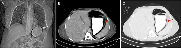
Gastric pneumatosis, a rare condition characterised by the presence of air in the gastric wall, may be caused by infection or ischaemia.1 A 47‐year‐old woman who had undergone a Hartmann procedure for sigmoid colon perforation, developed sepsis a week after surgery. Blood cultures grew Corynebacterium diphtheriae and Pseudomonas aeruginosa. A computed tomography scout image of the abdomen showed circumferential gas within the gastric wall (Figure, A). Axial images in soft tissue (Figure, B) and lung windows (Figure, C) delineated the extent of gastric pneumatosis. There was no evidence of portal venous gas. Gastric pneumatosis can be subdivided into gastric emphysema (typically self‐limiting) or the more aggressive emphysematous gastritis.2


  • Siti Soraya Ab Rahman1
  • Mohamad Farhan Mohamad Yusoff1

  • Universiti Sains Islam Malaysia, Nilai, Malaysia


Correspondence: soraya@usim.edu.my


Patient consent:

The patient gave written consent for publication.


Competing interests:

No relevant disclosures.

  • 1. Tagliaferri A, Melki G, Mohamed A, et al. Gastric pneumatosis in immunocompromised patients: a report of 2 cases and comprehensive literature review. Radiol Case Rep 2023; 18: 1152‐1155.
  • 2. Schattner A, Glick Y. Gastric pneumatosis and its varied pathogenesis. QJM 2020; 113: 747‐748.

Author

remove_circle_outline Delete Author
add_circle_outline Add Author

Comment
Do you have any competing interests to declare? *

I/we agree to assign copyright to the Medical Journal of Australia and agree to the Conditions of publication *
I/we agree to the Terms of use of the Medical Journal of Australia *
Email me when people comment on this article

Online responses are no longer available. Please refer to our instructions for authors page for more information.