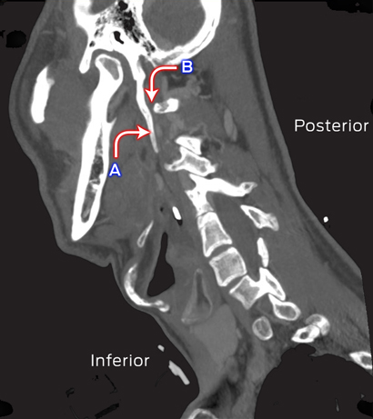
A 38-year-old man presented with influenza A (H1N1) pneumonia complicated by acute respiratory distress syndrome, resulting in an extended intensive care unit stay (37 days). After he was extubated, he was found to have left-side IX, X and XII cranial nerve palsies. Computed tomography of his neck showed bilateral elongated styloid processes (5.0 cm long; Figure), consistent with Eagle syndrome.1 The left styloid process was closely opposed to the transverse process of the first cervical vertebra, causing effacement of the internal jugular vein and compression of the IX, X and XII cranial nerves, which converge in this area. The eponym used to describe concurrent paralyses of the X and XII cranial nerves is Tapia syndrome,2 with prolonged neck flexion potentially contributing to the condition in our case.

Sagittal computed tomography image of the neck of the patient. A: Elongated styloid process (left); B: Area of compression of IX, X and XII cranial nerves.
Provenance: Not commissioned; externally peer reviewed.


Fraser Brims (Respiratory Department, Sir Charles Gairdner Hospital, Perth, WA) was the consultant who saw this patient.
No relevant disclosures.