


Clinical record
A 65‐year‐old man was referred to a private dermatologist for diagnosis and management of a hyperkeratotic, scaly, pruritic eruption affecting the trunk and extremities, which had been worsening over 12 months. Moderate potency topical corticosteroids had failed to improve his condition. He reported a history of previously treated pulmonary tuberculosis, prior venous thromboembolism, and asthma–chronic obstructive pulmonary disease overlap syndrome. He denied using regular prescription or over‐the‐counter medications. He migrated to Australia from South America in 1969 and resides with his partner, who prepares his meals. After becoming unemployed 12 months ago, he struggled with persistent low mood and poor appetite. He smoked marijuana and denied significant alcohol consumption.
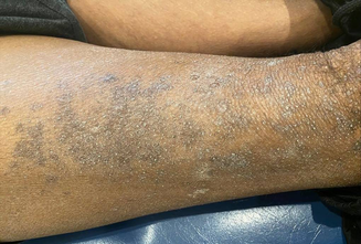
On examination, a keratotic folliculocentric, violaceous, scaly eruption affecting the trunk and extremities was observed (Box 1). Roughened nail plates and sparse brittle hair on the upper and lower extremities were noted. Dermoscopy revealed hyperkeratotic papules with prominent keratin plugging and white perifollicular scale (Box 2).
Based on these findings, the differential diagnosis consisted of primary follicular keratotic disease, and follicular variants of lichen planus, eczema and psoriasis. Results from a shave biopsy showed epidermal hyperkeratosis with follicular plugging and a perifollicular lymphocytic infiltrate. Blood results were remarkable for severe nutritional deficiencies of vitamin A (<0.4 μmol/L; reference interval [RI], 0.7–3.0 μmol/L), vitamin D (23 nmol/L; RI, 50–140 nmol/L), and active vitamin B12 (24 pmol/L; RI, >35 pmol/L), the latter of which had contributed to macrocytosis. The results from an infectious disease screen were negative for hepatitis B, hepatitis C, human immunodeficiency virus, syphilis, strongyloidiasis and tuberculosis. A diagnosis of phrynoderma was confirmed.
After identifying the nutritional deficiency, the general practitioner counselled the patient on increasing his dietary intake and he was started on several supplements for vitamin A, D and B complex. He also received a topical keratolytic agent and twice weekly sessions of narrowband UVB phototherapy. At the six‐week follow‐up, the appearance of his skin had improved and he reported improved appetite.
Discussion
Malnutrition is thought to be rare in high income countries. Phrynoderma is classically associated with vitamin A deficiency but may also be seen with deficiencies of vitamin B complex, vitamin E and essential fatty acids, as well as in patients with severe malnutrition.1 Phrynoderma is now thought to be more closely associated with general malnutrition, rather than isolated vitamin deficiencies. These deficiencies may arise due to inadequate dietary intake, malabsorption, increased metabolic demand, or impaired use. In developed countries, phrynoderma is seen in individuals with fat malabsorption secondary to inflammatory bowel disease, coeliac disease, short bowel syndrome, or after bariatric surgery. Other susceptible population groups include those with anorexia nervosa, those following fad diets, and older or disadvantaged people who live alone.2,3
The predominant cutaneous findings of phrynoderma include symmetrically distributed, variable sized, follicular‐based papules with conical keratotic plugs that favour the extensor surfaces of the extremities.4 In more severe or prolonged cases, other locations may become progressively affected, last of which is the face. Other cutaneous findings include generalised xerosis, diffuse hyperpigmentation, and sparse fragile hair. Systemic manifestations include blepharitis, night blindness, diarrhoea, muscle weakness and neuritis. Severe vitamin A deficiency is associated with devastating eye disease, including conjunctival and corneal xerosis, Bitot spots and keratomalacia.
Diagnosis of phrynoderma is primarily clinical, based on the characteristic appearance of follicular papules or plugs in a patient with compatible nutritional deficiencies and associated features. Biopsy can be helpful to exclude other differential diagnoses. Evaluation of the serum levels of vitamin A (retinol), essential fatty acids (such as linoleic acid), and other relevant micronutrients (zinc, selenium, vitamin B complex) can help identify specific deficiencies. Serum retinol‐binding protein levels can be measured as an indirect indicator of vitamin A deficiency.
Affected patients benefit from multidisciplinary care, particularly in cases of severe malnutrition, with the types of care dependent on the presentation. Patients should be evaluated for any underlying disorder that may be responsible for the nutritional deficiency.
Initial treatment consists of addressing the underlying deficiencies through improving nutrition and micronutrient supplementation, often with high doses. Caution should be employed due to the risk of refeeding syndrome with malnourished patients, and hepatotoxicity with supraphysiological supplementation of vitamin A. After restoration of nutrition, skin lesions should resolve over one to four months. Symptomatic relief may be achieved using gentle topical keratolytic agents, such as 10% urea in a cream base in addition to emollients. Phototherapy helps improve the symptoms and restore the vitamin D levels.
This case emphasises the importance of recognising cutaneous signs of nutritional deficiencies and undertaking appropriate investigations to promptly treat with suitable supplements.
Lessons from practice
- Phrynoderma is more closely associated with general malnutrition, rather than isolated vitamin deficiencies (eg, vitamins A, B complex, C or E).
- Consider the diagnosis of phrynoderma for any scaly, follicular‐based eruption, in at‐risk patients.
- A comprehensive assessment should be undertaken to determine the contributors to the malnourished state, including underlying medical conditions and psychosocial factors.

Provenance: Not commissioned; externally peer reviewed.
- 1. Bleasel NR, Stapleton KM, Lee MS, Sullivan J. Vitamin A deficiency phrynoderma: due to malabsorption and inadequate diet. J Am Acad Dermatol 1999; 41: 322‐324.
- 2. Di Stefani A, Orlandi A, Chimenti S, Bianchi L. Phrynoderma: a cutaneous sign of an inadequate diet. CMAJ 2007; 177: 855‐856.
- 3. Chia MW, Tay YK, Liu TT. Phrynoderma: a forgotten entity in a developed country. Singapore Med J 2008; 49: e160‐2.
- 4. Maronn M, Allen DM, Esterly NB. Phrynoderma: a manifestation of vitamin A deficiency?…The rest of the story. Pediatr Dermatol 2005; 22: 60‐63.
Open access:
Open access publishing facilitated by The University of Sydney, as part of the Wiley ‐ The University of Sydney agreement via the Council of Australian University Librarians.
Patient consent:
The patient provided written consent for publication.
No relevant disclosures.