


A 31-year-old woman presented with a 20-year history of lesions on the skin of her neck and axillae. Her family history was unremarkable.
Physical examination revealed multiple pinhead-sized yellowish flat-topped papules on the skin of the axillae and lateral aspects of the neck (Figure, A). These lesions made us suspect pseudoxanthoma elasticum, so we performed a fundoscopic examination which showed obvious angioid streaks (Figure, B). A biopsy of the axillary lesions showed a band of degenerated clumps of elastic fibres (Figure, C) and calcium deposits in the upper reticular dermis.
A diagnosis of pseudoxanthoma elasticum (PXE) was made. This condition is caused by mutations in the gene that encodes the ABCC6 protein, an ATP-binding cassette, subfamily C member.1
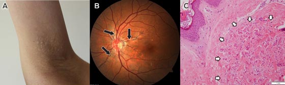
A: Photograph of the axilla of our patient showing several yellowish waxy papules.
B: Fundus of the left eye showing angioid streaks characteristic of pseudoxanthoma elasticum.
C: Histological examination of the skin biopsy showed fragmentation of elastic fibres in the upper reticular dermis.
- Zhenying Zhang1
- Xiaoming Liu1
- Dermatology, University of Hong Kong — Shenzhen Hospital, Shenzhen, Guangdong, China.
- 1. Pfendner E, Uitto J, Gerard GF, et al. Pseudoxanthoma elasticum: genetic diagnostic markers. Expert Opin Med Diagn 2008; 2: 63-79.