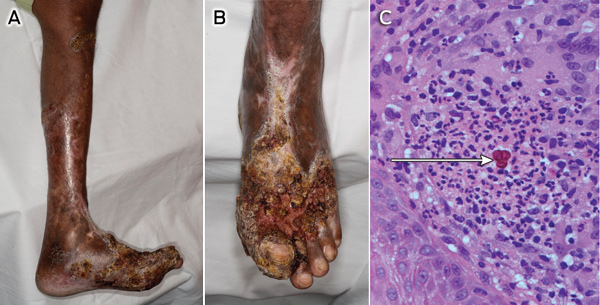
A 71-year-old farmer from the Solomon Islands presented in Melbourne with fungating skin lesions on his left lower leg. The process began 20 years earlier, leaving areas of scarring between the active sites (Figure, A and B). He was otherwise well.
A skin biopsy showed granulomatous inflammation with sclerotic bodies characteristic of chromoblastomycosis (Figure, C), and Fonsecaea pedrosoi was subsequently cultured and confirmed by ribosomal DNA internal transcribed spacer sequencing. Treatment combining surgical debridement, terbinafine and itraconazole is planned.
Although chromoblastomycosis occurs in the tropics worldwide, it is seldom reported in Pacific Island nations.1 Advanced presentations such as this are not uncommon in endemic areas.2
- 1. Woodgyer AJ, Bennetts GP, Rush-Munro FM. Four non-endemic New Zealand cases of chromoblastomycosis. Australas J Dermatol 1992; 33: 169-176.
- 2. Queiroz-Telles F, Esterre P, Perez-Blanco M, et al. Chromoblastomycosis: an overview of clinical manifestations, diagnosis and treatment. Med Mycol 2009; 47: 3-15.


We are grateful to Karin Leder and Robert Commons for their assistance with the manuscript, and to the microbiology staff of the Royal Melbourne Hospital, the Austin Hospital and Westmead Hospital for their support.